Advancements in Custom Cranioplasty: From CT Scans to Precision Implants
Cranioplasty, the surgical repair of skull defects, has undergone a significant technical evolution over the past two decades. What was once largely dependent on intraoperative improvisation and generic implant shaping is now anchored in preoperative digital planning and patient-specific device fabrication. For neurosurgeons and craniofacial surgeons managing large, irregular, or anatomically complex defects, this shift has direct implications for surgical efficiency, implant longevity, and patient outcomes.
This article examines the workflow behind modern custom cranioplasty, from initial imaging acquisition through implant delivery, with particular attention to the design considerations that distinguish high-fit, patient-specific implants from off-the-shelf alternatives.
The Role of CT Imaging in Implant Planning
The foundation of any patient-specific cranial implant is a high-resolution CT scan of the skull. Thin-slice axial imaging (typically acquired at 1 mm intervals or finer) generates the volumetric dataset from which implant geometry is derived. This data captures the contour of the existing cranium, the margins of the defect, and any relevant underlying anatomy.
Translating raw DICOM data into an implant geometry requires more than automated segmentation. Software-based reconstruction provides a baseline model, but artifacts from prior hardware, bone fragment irregularities, or previous cranioplasty mesh can introduce distortions that automated tools do not reliably resolve. This is one reason that many specialized implant manufacturers incorporate a review step (often involving experienced medical modelers or trained medical artists) to refine digital geometry before production begins.
The goal at this stage is not simply to fill the void but to restore the natural curvature and thickness of the surrounding skull. An implant that achieves anatomical contour continuity reduces palpable edges, supports overlying soft tissue, and minimizes the mechanical stress concentrations that can occur at implant-bone interfaces.
From Digital Model to Physical Device
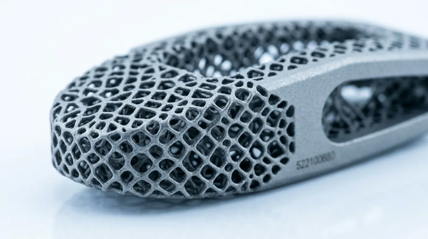
Once the implant geometry is validated, fabrication can proceed through one of several manufacturing pathways depending on the chosen material and the complexity of the case. Computer-aided design (CAD) files are typically used to drive CNC milling, additive manufacturing processes, or vacuum-formed sheet material shaping.
Polymethylmethacrylate (PMMA) has been one of the most widely used materials for custom cranial implants due to its biocompatibility, radiolucency, ease of intraoperative modification, and established clinical record. Patient-specific PMMA implants can be pre-formed to match the CT-derived geometry, eliminating the need for surgeons to hand-mix and shape the material intraoperatively, a process that carries risks of thermal injury and dimensional imprecision.
Porous polyethylene (PPE), titanium mesh, and hydroxyapatite-based composites each offer distinct mechanical and biological profiles. Titanium provides high structural rigidity useful in load-bearing frontal and temporal locations. Hydroxyapatite composites are osteoconductive, supporting potential bone ingrowth in select patient populations. The material selection is ultimately a clinical decision, shaped by defect location, patient age, prior surgical history, and surgeon preference.
The “Drop-In Fit” Objective
A consistent benchmark in patient-specific cranioplasty is achieving what practitioners sometimes describe as a “drop-in fit”, an implant that seats accurately within the defect margins without requiring significant intraoperative adjustment. This outcome depends on the precision of both the imaging acquisition and the fabrication process.
Defect margins that are irregular, beveled, or partially occluded by existing hardware present the greatest fitting challenges. Cases involving bilateral craniotomies, where the defect spans the midline, require careful attention to bilateral symmetry. Frontal bone defects that extend into orbital or nasal regions introduce complex three-dimensional geometry that flat or generically shaped implants cannot address.
For these high-complexity scenarios, manufacturers who invest in manual refinement steps alongside digital tooling tend to produce implants with more reliable fit characteristics. A technically precise implant reduces operative time, decreases the risk of implant migration or rocking, and supports a more predictable closure.
Preoperative Planning and Surgical Workflow Integration
Beyond the physical implant, custom cranioplasty workflows increasingly support preoperative surgical planning tools. Physical or 3D-printed skull models derived from the same CT dataset allow the surgical team to rehearse implant placement, confirm orientation, and identify potential interference points before the patient enters the operating room.
This integration of the implant design process with the surgical plan represents a broader shift toward procedural efficiency in reconstructive cranial surgery. Reducing intraoperative uncertainty in complex cranioplasty cases has documented implications for operative duration, blood loss, and anesthetic risk, particularly relevant for patients who have already undergone multiple prior procedures.
Quality and Regulatory Considerations
Patient-specific cranial implants in the United States are regulated by the Food and Drug Administration as Class II or Class III devices depending on their material and indication. Custom devices may be fabricated under the FDA’s Custom Device Exemption (CDE), which permits manufacture of a device that differs from a cleared predicate to meet the unique needs of an individual patient, subject to specific documentation and patient acknowledgment requirements.
Manufacturers operating under the CDE are not required to obtain 510(k) clearance for each custom implant, but they remain subject to FDA Quality System Regulation requirements, including design controls, process validation, and complaint handling. Surgeons working with custom implant providers should verify that their manufacturing partner maintains appropriate regulatory compliance documentation.
Summary
Custom cranioplasty today is a tightly coordinated process linking diagnostic imaging, computational design, skilled fabrication, and surgical execution. The shift from generic implants to patient-specific devices has expanded the range of defects that can be addressed with predictable outcomes. As imaging resolution and manufacturing precision continue to improve, the clinical standard for what constitutes an acceptable cranioplasty result continues to rise.
For further reading on complex cranial defect management and implant selection, see the related articles on bilateral craniotomy reconstruction and the role of medical artistry in implant design.